CONTENT WARNING – GRAPHIC IMAGERY
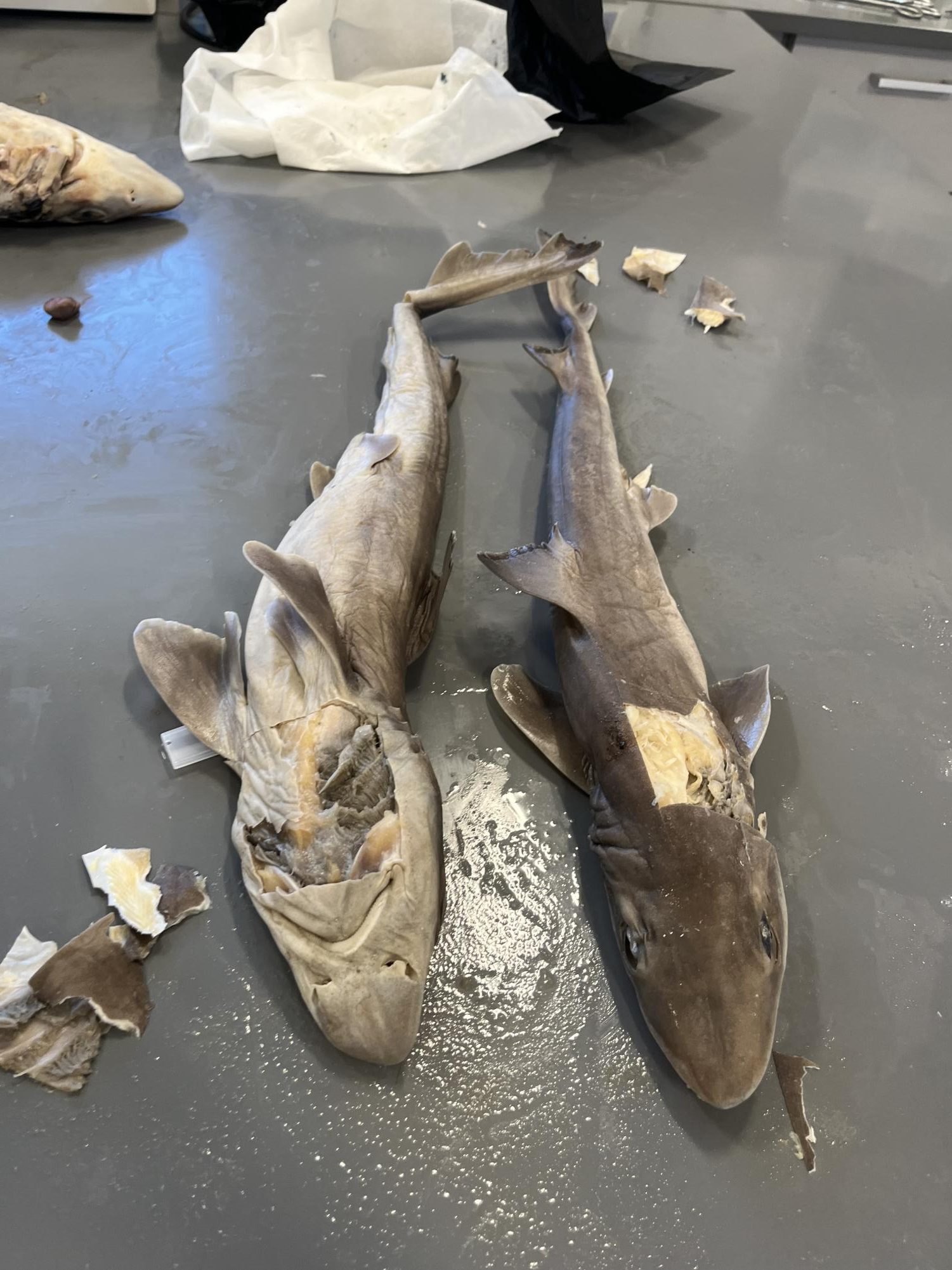
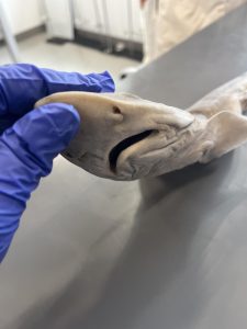
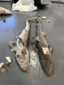
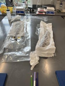
Throughout the week starting on April 8th, students in Dr. Evangelista’s biology class were given the opportunity to dissect dogfish sharks (genus squalus). This dissection consisted of two days of an in-depth hands-on experience where students were able to learn about both the external and internal anatomy of these sharks.
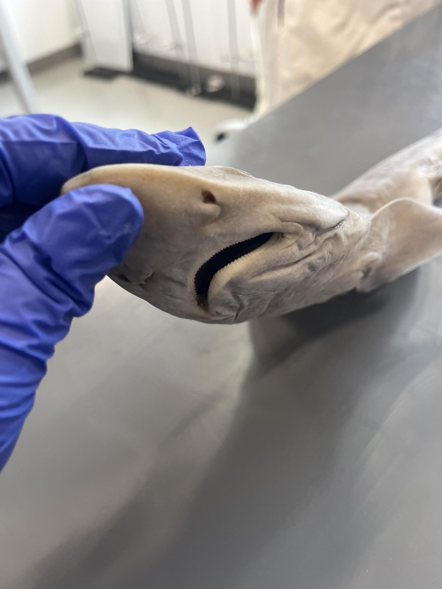
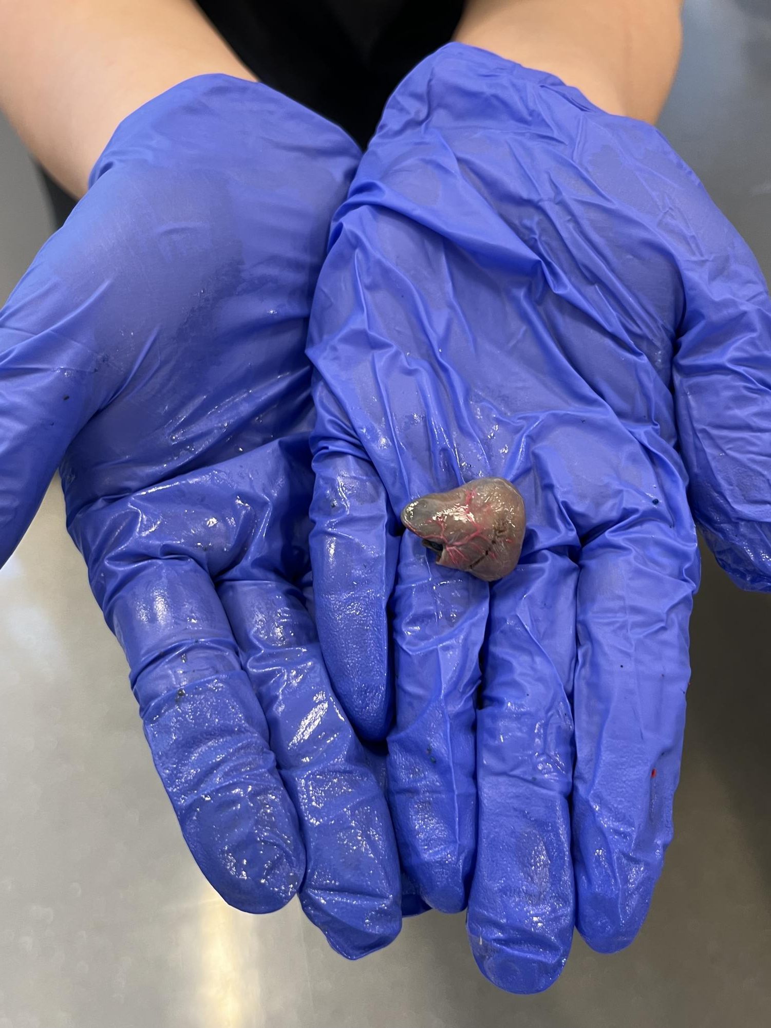
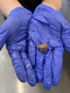
On day one, students examined the external anatomy, focusing on appendages, external sensory structures, skin, scales, eyes, membranes, mouth, and nares. On day two, students spent time examining the internal anatomy, including the hypaxial and epaxial musculature, buccal and branchial cavities, the spinal cord, the brain, heart and gills, and visceral organs. Dr. Evangelista said, “first-hand observations of natural structures in organisms are an important part of biology […] especially for students motivated towards medicine and life sciences; but also for general science knowledge and for students only just discovering their interests in biology.”
Students were able to complete the dissection at their own pace, allowing them to stay within their comfort level. Dr. Evangelista allowed students to take walks or breathers, and for those with religious or moral reasons, he allowed them to complete the same observations of form and functions by working with plants instead. Meera Iyer ‘25 said, “I was extremely excited that my class was going to do hands-on work.” But dissecting once living organisms is not for the faint of heart. Once the dissection began, Iyer said, “the odor in the classroom definitely had me on the fence but seeing Dr. Evangelista describing the various unique parts of each fish displayed fascinated me and encouraged us all to participate.” This hands-on dissection experience not only deepened students’ understanding of living organisms, but also sparked curiosity and passion for potential further exploration in the field.





Dr E • May 22, 2024 at 2:43 pm
Sophia Greller is the best reporter ever